Tuesday, January 31, 2017

pGLO Lab

1.
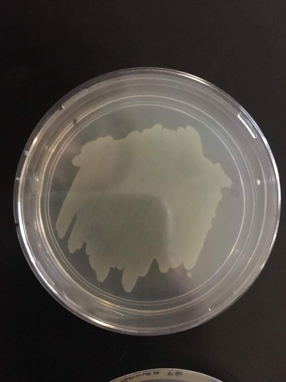
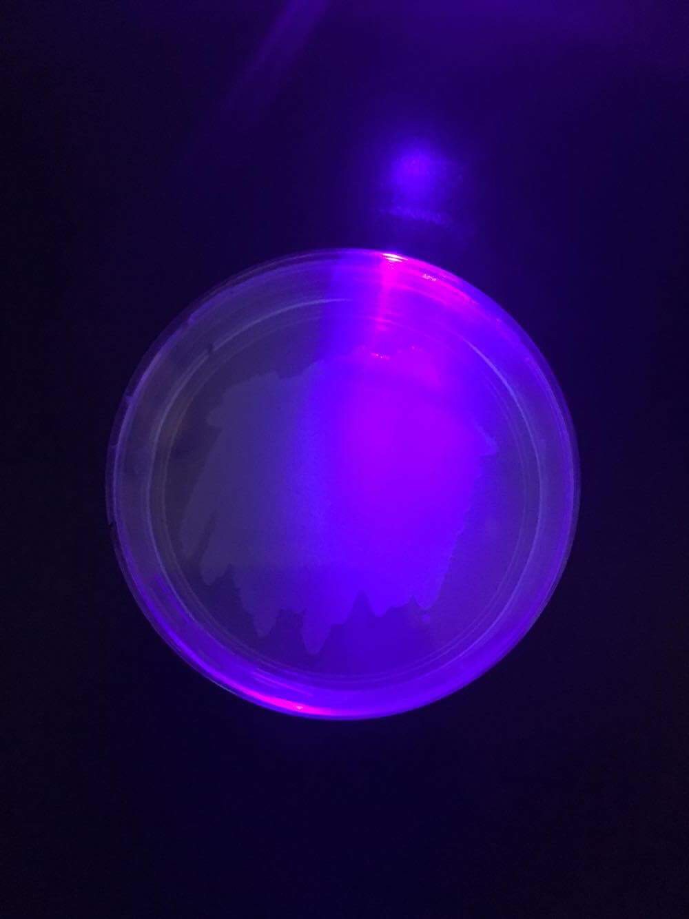
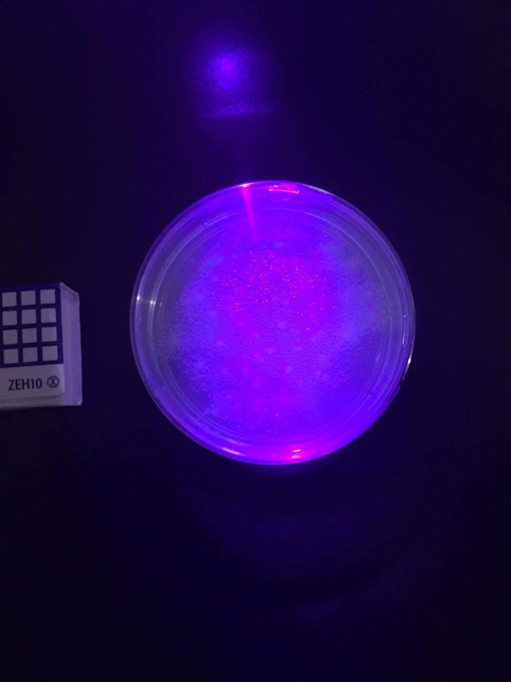
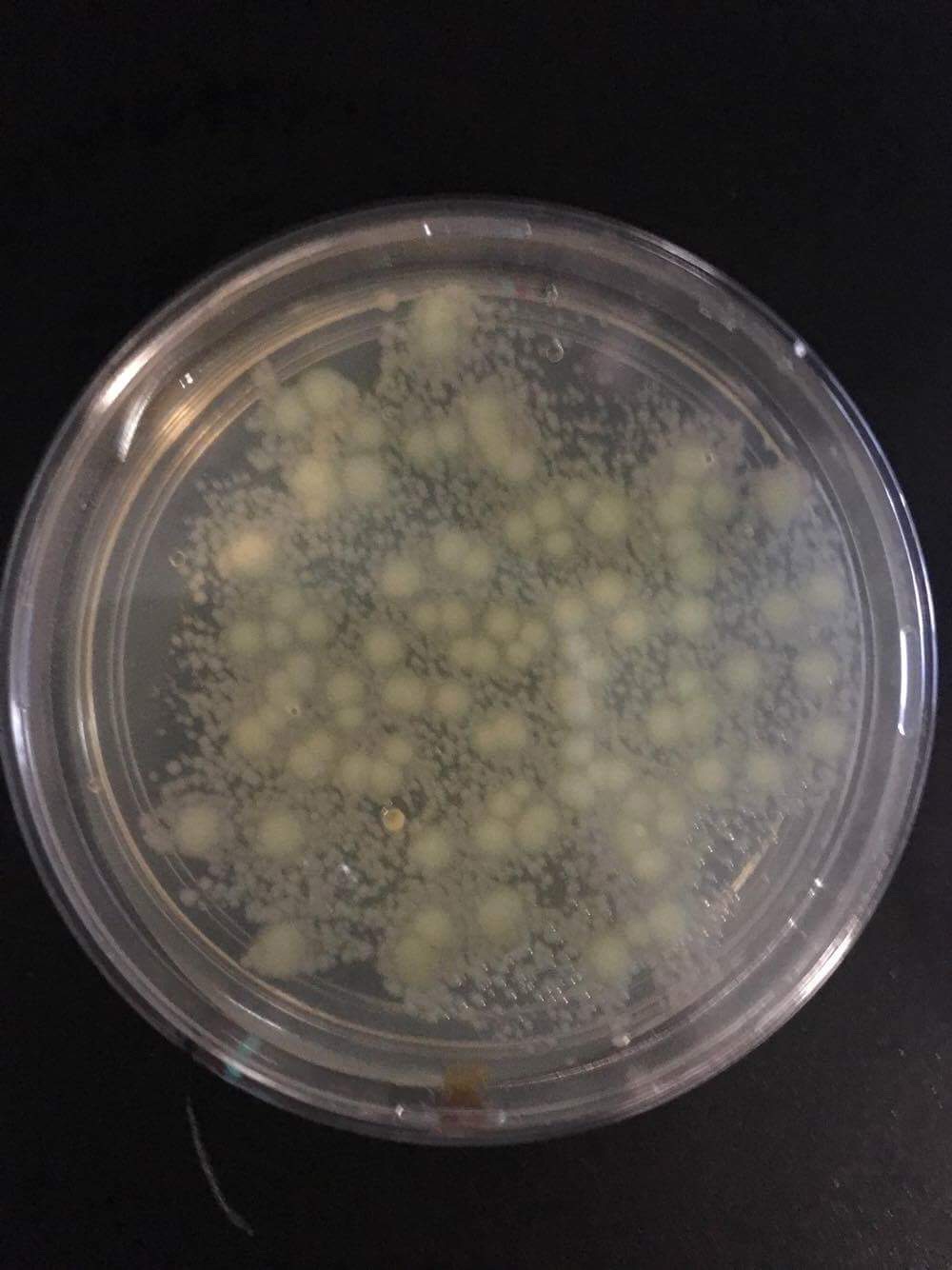
Obtain your team plates.  Observe your set of  “+pGLO” plates under room light and with UV light.  Record numbers of colonies and color of colonies. Fill in the table below.

Plate
Number of Colonies
Color of Colonies Under Room Light
Color of Colonies Under UV Light
-pGLO LB
0
Tan
Purple (the color of UV light)
+pGLO LB/amp
About 58
Tan
Purple (the color of UV light)
+pGLO LB/amp/ara
About 99
Tan
Green

2.
What two new traits do your transformed bacteria have?
The transformed bacteria glows green under the UV light and the bacteria is now resistant to the antibiotic ampicillin.

3.
Estimate how many bacteria were in the 100 uL of bacteria that you spread on each plate. Explain your logic.

An E. Coli cell is about 2 micrometers cubed and 1 micrometer is equal to (1e+9), which is 1,000,000,000. Since we spread 100 microliters of bacteria onto each plate, there were 100 times 1,000,000,000 bacteria on the plate, equalling a total of 100,000,000,000 bacteria that we spread on each plate.
4.
What is the role of arabinose in the plates?
The role of the arabinose was to help the bacteria glow and to actually activate the pGLO plasmid. As was stated in the vodcast, the protein GFP is supposed to make the bacteria glow, but in order for the GFP to be activated, the arabinose is used to trigger that protein, making the bacteria glow..
5.
List and briefly explain three current uses for GFP (green fluorescent protein) in research or applied science.
  • GFP can be used to serve as a marker protein, where when it attaches to and mark another protein, the scientists are then able to see the presence of that protein.
  • GFP can also be used to study bacteria more easily.
  • GFP can also be used to study diseases like HIV and track the spreading of those diseases.
6.
Give an example of another application of genetic engineering.

Genetic engineering has many applications in the medical field. One of the earliest applications of genetic engineering in pharmaceuticals was gene splicing to mass produce insulin in the body.


IMG_7078.JPG
-pGLO LB without UV light
IMG_7073.JPG
-pGLO LB with UV light


IMG_7080.JPG
+pGLO LB/amp without UV light


IMG_7075.JPG
+pGLO LB/amp with UV light


IMG_7081.JPG
+pGLO LB/amp/ara without UV light


IMG_7076.JPG
+pGLO LB/amp/ara with UV light

Thursday, January 19, 2017

Candy Electrophoresis Lab


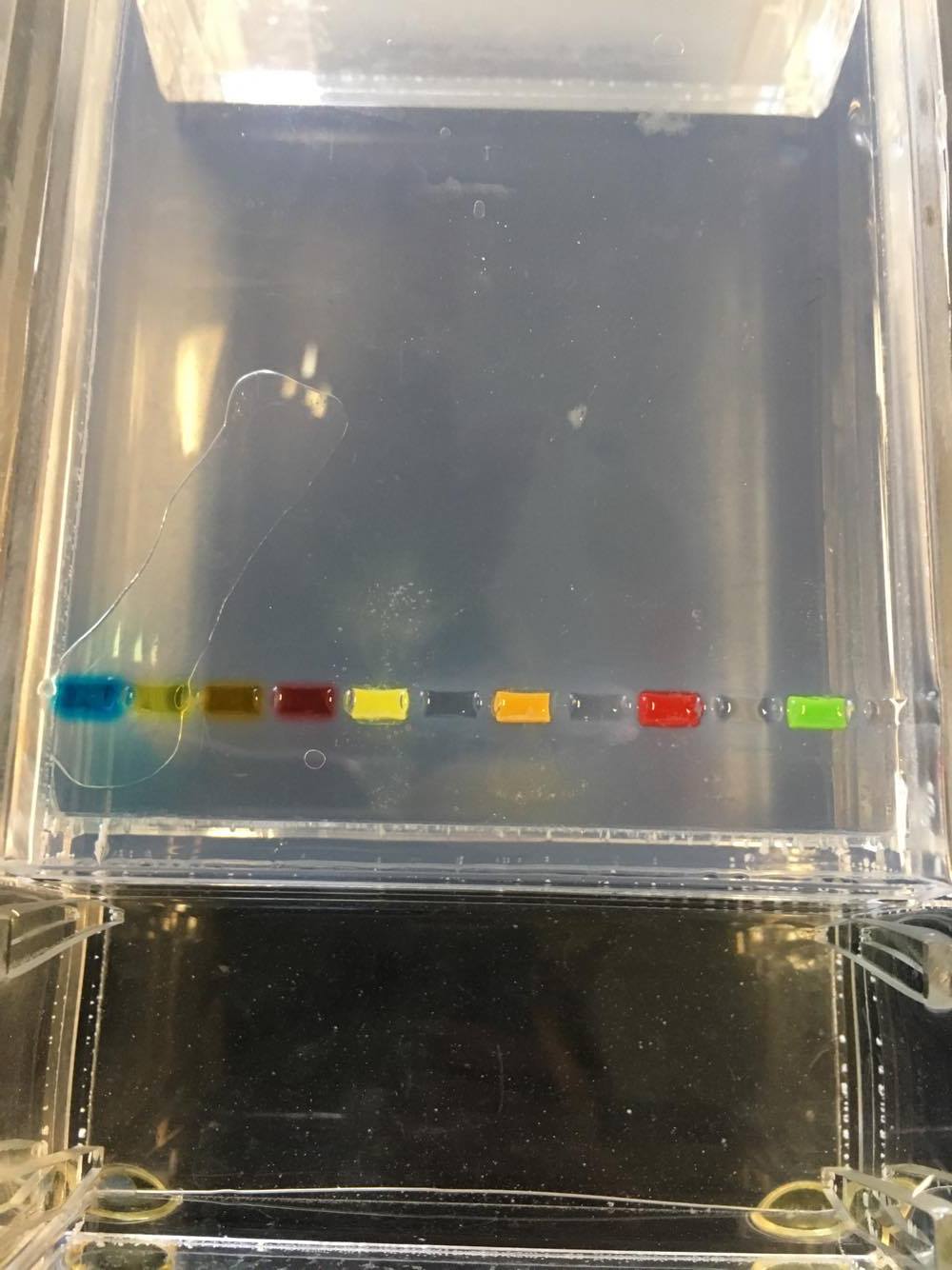
In this lab, we used the process called Gel Electrophoresis to observe the different colors of the candies. Comparing the four reference dyes to the four experimental sample, they acted similarly, meaning they moved at about the same rate and the bands were about the same size. In addition, all of the dyes moved in the “right” direction, from the negative end to the positive end of the gel.
The four on the left were the reference dyes and the four on the right were the experimental samples.
Just_dyes_bio_before_experiment.jpg
These were the samples after the process. The 3 yellow bands were at about the same length and were the same color. The 2 red bands, similarly, were the same length and their colors were almost identical. Like the yellow and red bands, the 2 orange bands looked very similar. However, the green experimental sample seemed to split into two separate bands, one yellow, which acted similar to the yellow reference dye, and one blue, which was the same length but a lighter color than the blue reference dye.
Just_Dyes_after_experiment_bio.jpg
Looking at the 4 dye structures, Betanin, Carminic Acid, Fast Green FCF, and Citrus red 2, Fast Green FCF has the same structure as the blue reference dye, which is why the Fast Green FCF would react similarly to the blue reference dye. Since the structures with the longest length move the slowest, and in this experiment the blue dye moved the slowest, structures with lengths longer than Fast Green FCF wouldn’t be comparable to the dyes from this experiment, which is why Betanin wouldn’t act similar to any of the dyes from this experiment. In this experiment, the blue dye moved the slowest out of the other dyes by a noticeable amount. Since Carminic Acid has a structure similar but not identical to Fast Green FCF, and the blue dye from the experimental sample, which came from skittles, had a structure similar but not identical to the blue dye, the Carminic Acid would be comparable to the blue band that split out of the green experimental sample. In addition, since the shorter structured band travels the fastest and the structure is similar to the yellow reference dye, the Citrus red 2 would be comparable to the yellow bands from the experiment.
Artificial food coloring is put in many foods including those for dogs as a strategy to make the food appear more appealing. The foods with a more appealing look would be bought by more people, making the company more money but also encouraging them and other companies to add more artificial food coloring to be placed in dog food.
In the process of Gel Electrophoresis, the main factors that contribute to the migration of the dyes are their size, the electrical current passing through, and for how long we left the electrical current passing through. The smaller the size the faster the dye passes through the gel as shown by the yellow dyes. However, as the longer dyes pass through much slower as shown by the blue dyes. In this experiment, we had the bands electrophoresed for 15 minutes at 100 volts. However, the main force that causes the process of Gel Electrophoresis to occur is the electrical current.
There are small holes in the agarose. Because of the size of the bands, the smaller structures are able to move through the gels more easily and thereby quicker. As a result of moving at a slower pace, the longer strands get left behind, which is how the molecules are able to separate by size. If the molecules with the molecular weight of 600, 1000, 2000, and 5000 daltons, I believe the molecules with the molecular weight of 600 would travel the farthest, the molecule with 1000 daltons would travel second fastest, the molecule with 2000 daltons would come in third, and the molecule with 5000 daltons would move the shortest distance.

Tuesday, January 10, 2017

New Year Goals

Throughout last semester, I saw myself wondering how 24 hours a day went by so quickly. I allotted 9 hours to school, 6 hours to sleep, and 3 hours to extracurricular activities per day. That would leave me with about 6 hours of homework and studying. Six hours is enough to review the day, finish my homework and any studying that would entail, and prepare for the next day. However that management of my time never truly came into action last semester due to not being able to manage my time as well as I could have, as I was struggling to sleep on time, I wasn’t focusing my full effort on the task that I was completing, be it studying for science or english or math, or even watching a vodcast. This semester I will be better at managing my time. I will put my full attention onto that vodcast that I will watch, or the textbook notes that I will complete. This would lead to me gaining a better understanding of the topic but also leaving me with ample time to finish the rest of the tasks at hand. This semester through managing my time better, I will make sure I am able to complete every task at hand without getting distracted and with my full attention, which would be measured by my improving school performance.
However, procrastination and being unable to manage time right come together. As a result of me not managing my time as well as I could have, it lead to me procrastinating on small things. However once you start to procrastinate, you end up getting caught in a never ending cycle until a solid break. This semester I will not procrastinate. The day I get assigned the vodcast is when I will finish it rather than leaving it off until the next day, the day before the class it’s due. And when we write our lab conclusions or unit reflections, I will work my hardest to finish it during class instead of leaving it until I get home, which leads to me struggling to turn in work that I know could have been better a minute before the 11:59 p.m. deadline. This semester will be the semester where I will not put off a single task until later. I will manage my time better, reducing my procrastination, which could also be measured by my improving school performance.

Thursday, December 15, 2016

Unit 5 Reflection

Throughout this unit, we learned about the Central Dogma, which is the many processes in which proteins are formed.
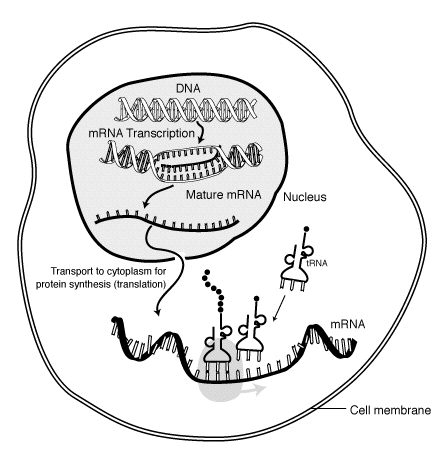
First the DNA unzips. Then the mRNA attaches to the unmatched bases. But as it attaches, it replaces the thymine with uracil. Then the mRNA detaches from the half of the DNA and the DNA zips back up. The mRNA then leaves the nucleus through the nuclear pores to go to the cytoplasm. That entire process is one part of protein synthesis and is called transcription.
Then, after the mRNA comes to the cytoplasm, it attaches to the ribosome, where it reads the bases in triplet groups called codons. The mRNA starts with the start codon signalling the ribosome to start reading it and the stop codon in the end signals the ribosome to stop reading the sequence. While the ribosome reads the sequence, the tRNA then brings the amino acids, which then attach to each codon. Eventually an amino acid is attached to each codon in the sequence where it then detaches from the mRNA and tRNA. This process is called translation. The mRNA is then sent back through the nuclear pores where it is recycled in the nucleus and the tRNA is sent back to the cytoplasm where it is recycled there as well.
amino-acids-metabolism-figure-2-149BBF6DDD969BAB1D1.png
Finishing off the protein synthesis process, the amino acids then bond together to form a protein. The protein is then sent off to the rough endoplasmic reticulum and then sent to the Golgi apparatus where it is then shipped out of the cell using vesicles to help the body function.
Unknown------------666.jpg
This unit really brought together all of the concepts from day one, because everything we started was finished, meaning that in a cell, we learned what each of the organelles do in previous units. Then we saw how the organelles contribute to the creation of proteins and how proteins are necessary. We then finished off with how proteins are made specifically and how after the mRNA and tRNA is recycled and the finished proteins are sent back to be used by the body. This unit really brought everything full circle.
However although I say that this unit really brought everything full circle, it took me a while to really understand how this related to what else we learned in this unit, which was one of the struggles that I had to face throughout this unit. But by using what I learned in the VARK Questionnaire that I took last unit, I learned that diagrams and visuals were helpful to me, which is why by making diagrams and writing out the whole process I was able to connect the concepts.
I struggled with wondering with what happens next. For example, I wondered where exactly tRNA came from but also where it goes after the amino acids bond.
But my strengths came after my initial struggle. My being able to understand how everything came together was a strength because it helped me understand the concepts better.
I still want to know more about RNA. During this unit it was brought up that a ribosome actually is just a bunch of RNA, which is a little bit hard to grasp still.
I consider myself a better student than I was in the previous units, because I learned how to use my strengths to my advantage. Since I am more of a visual person, I drew out all of the diagrams and wrote out all of the processes. I know how to study better, which is a skill that I can use not only in the science class but outside it as well.

Wednesday, December 14, 2016

Protein Synthesis Lab

In this lab, we asked the question, “How does the body produce proteins?” In the protein synthesis process, there are two steps — transcription and translation. Transcription occurs first, where one strand of the DNA is copied and makes RNA. Then the base thymine is replaced with uracil. After, the RNA is transported to the cytoplasm, where translation occurs. The ribosome in the cytoplasm reads the RNA in groups of three, or codons, where it forms amino acids. The amino acids then bond together to make proteins.
MRNA-interaction.png
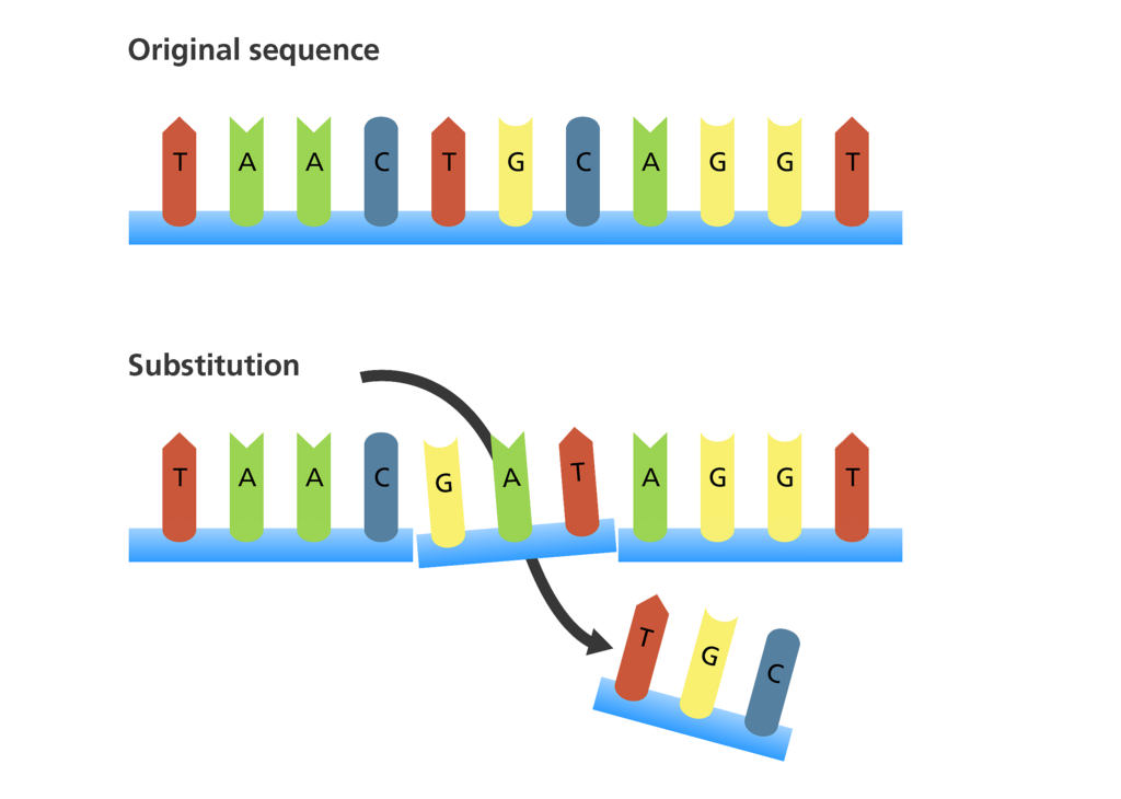
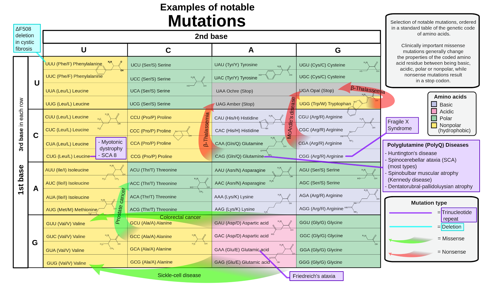
Changing bases in the DNA molecule result in either frameshift mutations or substitution. The frameshift mutations include insertion or deletion where a base is either added or deleted, and substitution replaces a base for another. In this lab, I noticed that the frameshift mutation had the most amount of change comparing it to substitution because by adding or deleting a base, the entire sequence is shifted and therefore changed, but in substitution, the one base that is being substituted is the only one being affected, so it wouldn’t affect the sequence by a great deal. However, the mutations are the most problematic and harmful when they occur at the beginning of the sequence, because the start codon could be changed, due to mutations, into the stop codon, making the amino acids unable to form into a protein.
26855220782_ff1e86c474_b.jpg
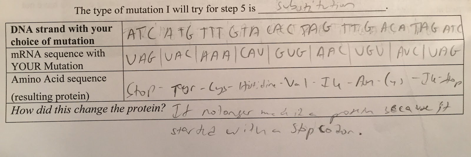
In the experiment, I observed how the substitution mutation would affect the sequence. I substituted the start codon for a stop codon, which invalidated the protein because the protein was never able to form as it started with a stop codon, signalling the stopping of the process. Although this mutation greatly affected the protein, it was only because of where it occurred, rather than how the mutation occurred. Since only the first three bases were substituted, nothing happened to the rest of the codons as they stayed the same. But due to where the mutation was placed, it affected the protein, as the mutation I tested substituted the start codon for a stop codon.
FullSizeRender-3.jpg
Mutations are so common in life. However these mutations can have dramatic effects that can put the individual at great risk, or it can barely affect that individual at all. For example, the fatal disease called Tay-Sachs disease comes from the frameshift mutation on the gene that converts the alpha-subunit of the lysosomal enzyme, beta-hexosaminidase. This frameshift mutation is the cause of Tay-Sachs disease and cause the destruction of the nerve cells in the spinal cord and the brain, which has been shown to be fatal.

Notable_mutations.png

Tuesday, December 6, 2016

Human DNA Extraction Lab

In this lab, we asked the question, “How can DNA be separated from cheek cells in order to study it?” I found that after the cold isopropanol alcohol was added to the solution, the DNA was separated. In order for the DNA to be separated from my cheek cells, I homogenized the cell tissue with the gatorade and the saliva, I washed the solution out with soap during the lysis process, then I added the salt and the pineapple juice facilitated the precipitation process as they broke down the remaining histones, and lastly to separate the DNA from the cell, I added the cold isopropanol alcohol to the solution. During the homogenization, lysis, and precipitation processes, the cells started to become visible and eventually the individual DNA molecule started to become visible. The claim that the alcohol is what caused the DNA to separate from cheek cells could come from the knowledge that DNA is polar and the alcohol is nonpolar. Since the DNA separates after the alcohol is added because of the clash in the polarity, it would support my claim that after the cold isopropanol alcohol was added to the solution, the DNA was separated.
While my hypothesis stating that the alcohol will cause the DNA to separate was supported, there could have been errors. Some of the errors that occurred that could have affected the outcome was when after the pineapple juice was added, the mixing process led to some of the solution spilling out of the tube due to the ineffective cover that was placed on the test tube that could have led to some important molecules that affected the effect of the enzyme on the DNA extraction process. The second error that occurred had to do with the amount of alcohol that was put into the solution. Since I didn’t measure the exact amount of the alcohol and instead put an estimated amount of alcohol, it could have led to affecting the molecule of DNA. In order to prevent these mistakes in the future, I could use a better stopper for the tube that covers the tube in a way so none of the solution spills out. In addition, to prevent the harming of the DNA, I could measure the amount of solution I had and then measure exactly the same amount of alcohol to add to the solution instead of estimating the amount of alcohol. So mainly, both of my mistakes could be prevented by being more accurate.
This lab was done to show how DNA is extracted and the different visible stages in which the DNA goes through during the extraction process. This lab relates to what we’ve learned in class because currently we are studying DNA and its replication process, so through this lab, we were able to see the most basic of the processes, the DNA being extracted. This could be applied to future situations because if I go into a field of genetics, then DNA extraction along with far more complicated procedures would be what I would do.

Tuesday, November 29, 2016

Unit 4 Reflection

Throughout this unit, we answered the question, “Why is sex so great?” During this unit we did the Coin Sex Lab as a means to predict the different possibilities of the traits for the hypothetical offspring, by flipping coins. The coins served as a model for genetic concepts because it shows how the acquiring of traits doesn’t stick to a formula. It shows the random nature of genetics and the acquiring of genes.Then we compared the probabilities of the possibilities from the punnett squares to the traits that we got as a result of flipping the coins. We experimented with both hybrid and dihybrid crosses. In the dihybrid cross simulation we were expecting the results of 9:3:3:1 for the genotype. In the end, we ended up with the results much different. I attribute these different results to the random nature of genetics because although any amount of punnett squares can get the probability of the possibilities, the main reason for our results was that genetics is random.
The randomness associated with genetics and acquiring different genes become a limitation of using probability to predict the offspring’s genes. The completion of punnett squares and even more extensive genetic testing can only get you so far, as genetics, although it has its patterns, is random.
This understanding of genetics and how it is acquired relates to my life because I now understand my specific genetics. It explains where my height, my eye color, my skin color, and my other traits from genetics, came from. My traits, as I have learned through this unit, didn’t only come from my parents but could have skipped a generations. It made me take a conscious look at my traits and left me wondering if there were any genes that could have been recessive in previous generations but appeared in my sisters or myself.
But although acquiring specific played a large role in this unit, this unit was about what is sex, what does sexual reproduction mean, and what comes as a result of sexual reproduction. With those main topics being covered, we learned about the difference between mitosis and meiosis. Mitosis is part of the cell cycle where the cell splits then duplicates into more cells. However, meiosis isn’t really a cycle. Meiosis is when the cell splits into 2 gametes or haploids. Then those 2 gametes split into 4 daughter cells, or gametes, during meiosis 2. Then these gametes can be fertilized, bringing it back to mitosis, where the cell cycle causes the cells to multiply and grow.
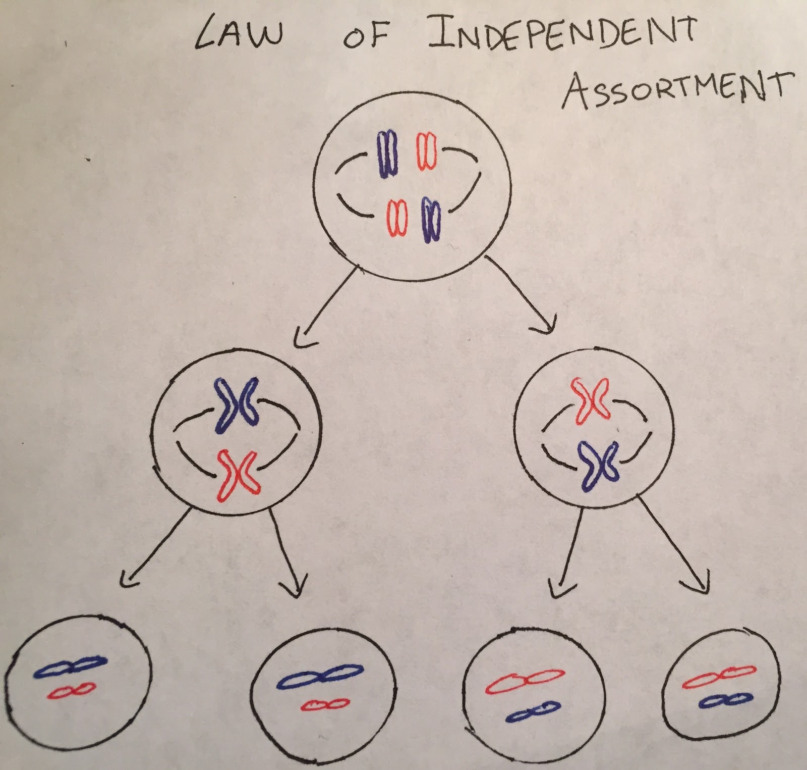
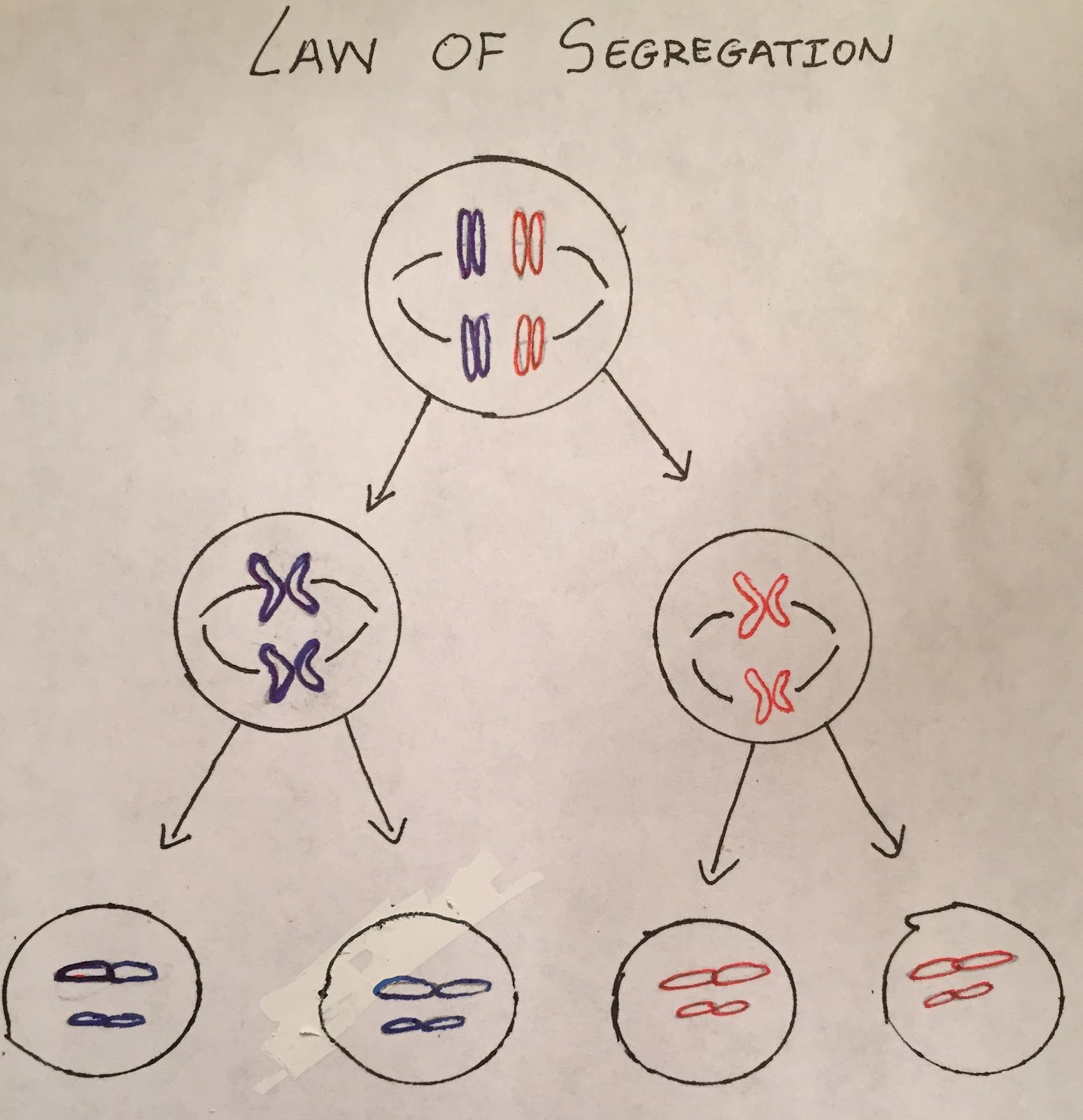
During meiosis, the genes are assorted through the law of independent assortment or the law of segregation.
Law of independent assortment.jpg                          Law_of_Segregation.jpg
In addition, we learned about genetics and the different patterns of genetics. We learned about the different types of dominance- dominance, incomplete dominance, and codominance. We also learned about different types of genetic disorders such as autosomal or sex-linked.
During this unit, the genetic aspects of the unit came to me pretty easily, because most of it had to do with being able to solve punnett squares. When looking at the phenotype or the genotype of the offspring of two heterozygous parents, either memorization or solving the punnett square would give you the answer. When looking at the probability of a son having hemophilia when his mother is a carrier of hemophilia but doesn’t have it herself and his father is also a carrier, it would be 50%. This can be solved through a simple punnett square.
Unknown-----------1.png
However, learning the process of meiosis was difficult. Because mitosis and meiosis have always been said together, trying to understand that meiosis wasn’t a cycle but instead was a way to make gametes took a lot of time, making it much harder than the rest of the concepts from this unit.
But during this unit, we were assigned a project where we made an infographic. Doing the infographic was very helpful in solidifying my understanding of these concepts because in addition to learning about the concept in the vodcast and then testing ourselves through the check for understanding quizzes, we practiced each of the concepts while making our infographic.
After watching the vodcasts and taking the CFU’s, I still didn’t understand the different types of dominances very well, specifically codominance and incomplete dominance. However after I did the infographic, after I searched up more graphics and explained what each of those specifically meant, I was very clear on the concept.
This taught me how to be a better student and learner through a lesson that can be applied to anything I do, which is to not stop my learning at the vodcasts and the CFU’s but to take that extra step to understanding the concept, whether it be watching another video about the concept or finding a picture explaining the concept.