Thursday, December 15, 2016

Unit 5 Reflection

Throughout this unit, we learned about the Central Dogma, which is the many processes in which proteins are formed.
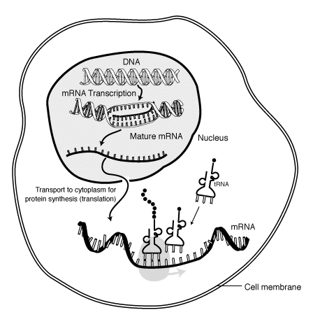
First the DNA unzips. Then the mRNA attaches to the unmatched bases. But as it attaches, it replaces the thymine with uracil. Then the mRNA detaches from the half of the DNA and the DNA zips back up. The mRNA then leaves the nucleus through the nuclear pores to go to the cytoplasm. That entire process is one part of protein synthesis and is called transcription.
Then, after the mRNA comes to the cytoplasm, it attaches to the ribosome, where it reads the bases in triplet groups called codons. The mRNA starts with the start codon signalling the ribosome to start reading it and the stop codon in the end signals the ribosome to stop reading the sequence. While the ribosome reads the sequence, the tRNA then brings the amino acids, which then attach to each codon. Eventually an amino acid is attached to each codon in the sequence where it then detaches from the mRNA and tRNA. This process is called translation. The mRNA is then sent back through the nuclear pores where it is recycled in the nucleus and the tRNA is sent back to the cytoplasm where it is recycled there as well.
amino-acids-metabolism-figure-2-149BBF6DDD969BAB1D1.png
Finishing off the protein synthesis process, the amino acids then bond together to form a protein. The protein is then sent off to the rough endoplasmic reticulum and then sent to the Golgi apparatus where it is then shipped out of the cell using vesicles to help the body function.
Unknown------------666.jpg
This unit really brought together all of the concepts from day one, because everything we started was finished, meaning that in a cell, we learned what each of the organelles do in previous units. Then we saw how the organelles contribute to the creation of proteins and how proteins are necessary. We then finished off with how proteins are made specifically and how after the mRNA and tRNA is recycled and the finished proteins are sent back to be used by the body. This unit really brought everything full circle.
However although I say that this unit really brought everything full circle, it took me a while to really understand how this related to what else we learned in this unit, which was one of the struggles that I had to face throughout this unit. But by using what I learned in the VARK Questionnaire that I took last unit, I learned that diagrams and visuals were helpful to me, which is why by making diagrams and writing out the whole process I was able to connect the concepts.
I struggled with wondering with what happens next. For example, I wondered where exactly tRNA came from but also where it goes after the amino acids bond.
But my strengths came after my initial struggle. My being able to understand how everything came together was a strength because it helped me understand the concepts better.
I still want to know more about RNA. During this unit it was brought up that a ribosome actually is just a bunch of RNA, which is a little bit hard to grasp still.
I consider myself a better student than I was in the previous units, because I learned how to use my strengths to my advantage. Since I am more of a visual person, I drew out all of the diagrams and wrote out all of the processes. I know how to study better, which is a skill that I can use not only in the science class but outside it as well.

Wednesday, December 14, 2016

Protein Synthesis Lab

In this lab, we asked the question, “How does the body produce proteins?” In the protein synthesis process, there are two steps — transcription and translation. Transcription occurs first, where one strand of the DNA is copied and makes RNA. Then the base thymine is replaced with uracil. After, the RNA is transported to the cytoplasm, where translation occurs. The ribosome in the cytoplasm reads the RNA in groups of three, or codons, where it forms amino acids. The amino acids then bond together to make proteins.
MRNA-interaction.png
Changing bases in the DNA molecule result in either frameshift mutations or substitution. The frameshift mutations include insertion or deletion where a base is either added or deleted, and substitution replaces a base for another. In this lab, I noticed that the frameshift mutation had the most amount of change comparing it to substitution because by adding or deleting a base, the entire sequence is shifted and therefore changed, but in substitution, the one base that is being substituted is the only one being affected, so it wouldn’t affect the sequence by a great deal. However, the mutations are the most problematic and harmful when they occur at the beginning of the sequence, because the start codon could be changed, due to mutations, into the stop codon, making the amino acids unable to form into a protein.
26855220782_ff1e86c474_b.jpg
In the experiment, I observed how the substitution mutation would affect the sequence. I substituted the start codon for a stop codon, which invalidated the protein because the protein was never able to form as it started with a stop codon, signalling the stopping of the process. Although this mutation greatly affected the protein, it was only because of where it occurred, rather than how the mutation occurred. Since only the first three bases were substituted, nothing happened to the rest of the codons as they stayed the same. But due to where the mutation was placed, it affected the protein, as the mutation I tested substituted the start codon for a stop codon.
FullSizeRender-3.jpg
Mutations are so common in life. However these mutations can have dramatic effects that can put the individual at great risk, or it can barely affect that individual at all. For example, the fatal disease called Tay-Sachs disease comes from the frameshift mutation on the gene that converts the alpha-subunit of the lysosomal enzyme, beta-hexosaminidase. This frameshift mutation is the cause of Tay-Sachs disease and cause the destruction of the nerve cells in the spinal cord and the brain, which has been shown to be fatal.

Notable_mutations.png

Tuesday, December 6, 2016

Human DNA Extraction Lab

In this lab, we asked the question, “How can DNA be separated from cheek cells in order to study it?” I found that after the cold isopropanol alcohol was added to the solution, the DNA was separated. In order for the DNA to be separated from my cheek cells, I homogenized the cell tissue with the gatorade and the saliva, I washed the solution out with soap during the lysis process, then I added the salt and the pineapple juice facilitated the precipitation process as they broke down the remaining histones, and lastly to separate the DNA from the cell, I added the cold isopropanol alcohol to the solution. During the homogenization, lysis, and precipitation processes, the cells started to become visible and eventually the individual DNA molecule started to become visible. The claim that the alcohol is what caused the DNA to separate from cheek cells could come from the knowledge that DNA is polar and the alcohol is nonpolar. Since the DNA separates after the alcohol is added because of the clash in the polarity, it would support my claim that after the cold isopropanol alcohol was added to the solution, the DNA was separated.
While my hypothesis stating that the alcohol will cause the DNA to separate was supported, there could have been errors. Some of the errors that occurred that could have affected the outcome was when after the pineapple juice was added, the mixing process led to some of the solution spilling out of the tube due to the ineffective cover that was placed on the test tube that could have led to some important molecules that affected the effect of the enzyme on the DNA extraction process. The second error that occurred had to do with the amount of alcohol that was put into the solution. Since I didn’t measure the exact amount of the alcohol and instead put an estimated amount of alcohol, it could have led to affecting the molecule of DNA. In order to prevent these mistakes in the future, I could use a better stopper for the tube that covers the tube in a way so none of the solution spills out. In addition, to prevent the harming of the DNA, I could measure the amount of solution I had and then measure exactly the same amount of alcohol to add to the solution instead of estimating the amount of alcohol. So mainly, both of my mistakes could be prevented by being more accurate.
This lab was done to show how DNA is extracted and the different visible stages in which the DNA goes through during the extraction process. This lab relates to what we’ve learned in class because currently we are studying DNA and its replication process, so through this lab, we were able to see the most basic of the processes, the DNA being extracted. This could be applied to future situations because if I go into a field of genetics, then DNA extraction along with far more complicated procedures would be what I would do.

Tuesday, November 29, 2016

Unit 4 Reflection

Throughout this unit, we answered the question, “Why is sex so great?” During this unit we did the Coin Sex Lab as a means to predict the different possibilities of the traits for the hypothetical offspring, by flipping coins. The coins served as a model for genetic concepts because it shows how the acquiring of traits doesn’t stick to a formula. It shows the random nature of genetics and the acquiring of genes.Then we compared the probabilities of the possibilities from the punnett squares to the traits that we got as a result of flipping the coins. We experimented with both hybrid and dihybrid crosses. In the dihybrid cross simulation we were expecting the results of 9:3:3:1 for the genotype. In the end, we ended up with the results much different. I attribute these different results to the random nature of genetics because although any amount of punnett squares can get the probability of the possibilities, the main reason for our results was that genetics is random.
The randomness associated with genetics and acquiring different genes become a limitation of using probability to predict the offspring’s genes. The completion of punnett squares and even more extensive genetic testing can only get you so far, as genetics, although it has its patterns, is random.
This understanding of genetics and how it is acquired relates to my life because I now understand my specific genetics. It explains where my height, my eye color, my skin color, and my other traits from genetics, came from. My traits, as I have learned through this unit, didn’t only come from my parents but could have skipped a generations. It made me take a conscious look at my traits and left me wondering if there were any genes that could have been recessive in previous generations but appeared in my sisters or myself.
But although acquiring specific played a large role in this unit, this unit was about what is sex, what does sexual reproduction mean, and what comes as a result of sexual reproduction. With those main topics being covered, we learned about the difference between mitosis and meiosis. Mitosis is part of the cell cycle where the cell splits then duplicates into more cells. However, meiosis isn’t really a cycle. Meiosis is when the cell splits into 2 gametes or haploids. Then those 2 gametes split into 4 daughter cells, or gametes, during meiosis 2. Then these gametes can be fertilized, bringing it back to mitosis, where the cell cycle causes the cells to multiply and grow.
During meiosis, the genes are assorted through the law of independent assortment or the law of segregation.
Law of independent assortment.jpg                          Law_of_Segregation.jpg
In addition, we learned about genetics and the different patterns of genetics. We learned about the different types of dominance- dominance, incomplete dominance, and codominance. We also learned about different types of genetic disorders such as autosomal or sex-linked.
During this unit, the genetic aspects of the unit came to me pretty easily, because most of it had to do with being able to solve punnett squares. When looking at the phenotype or the genotype of the offspring of two heterozygous parents, either memorization or solving the punnett square would give you the answer. When looking at the probability of a son having hemophilia when his mother is a carrier of hemophilia but doesn’t have it herself and his father is also a carrier, it would be 50%. This can be solved through a simple punnett square.
Unknown-----------1.png
However, learning the process of meiosis was difficult. Because mitosis and meiosis have always been said together, trying to understand that meiosis wasn’t a cycle but instead was a way to make gametes took a lot of time, making it much harder than the rest of the concepts from this unit.
But during this unit, we were assigned a project where we made an infographic. Doing the infographic was very helpful in solidifying my understanding of these concepts because in addition to learning about the concept in the vodcast and then testing ourselves through the check for understanding quizzes, we practiced each of the concepts while making our infographic.
After watching the vodcasts and taking the CFU’s, I still didn’t understand the different types of dominances very well, specifically codominance and incomplete dominance. However after I did the infographic, after I searched up more graphics and explained what each of those specifically meant, I was very clear on the concept.
This taught me how to be a better student and learner through a lesson that can be applied to anything I do, which is to not stop my learning at the vodcasts and the CFU’s but to take that extra step to understanding the concept, whether it be watching another video about the concept or finding a picture explaining the concept.

Monday, October 31, 2016

Is Sexual Reproduction Important?

Sex is a necessity when referring to the survival of organisms. As stated by Dr. Tatiana in Dr. Tatiana’s Sex Advice To All Creation by Olivia Judson, sex is the mixing of genes, creating an organism with a new genetic makeup. A new genetic makeup is essential for evolution to occur, and happens through mutation and sex. However some organisms evolve to reproduce asexually, or without sex. Although asexual reproduction has its benefits, it can lead to extinction rapidly, making sexual reproduction better and thereby more important.
In “Wholly Virgin,” many arguments were stated that supported the opinion that sex is important. The Philodina roseola was claimed to have been reproducing asexually for the past 85 million years without the need for meiosis. However, the ram pointed out that chaetonotoid gastrotrichs, who also claimed to be ancient asexuals, were caught making sperm, an activity complying with the sexual reproduction rather than asexual reproduction, making it more likely for more organisms claiming to be ancient asexuals to in fact reproduce sexually.
In addition, according to Muller’s ratchet, by geneticist Hermann Muller, “asexuals are evolutionarily short-lived because” (226). This theory shows that sexual reproduction is better because asexual reproduction will lead to an evolutionary halt which is detrimental to the organism.
Another benefit stemming from sexual reproduction, is the lack of exposure to diseases and a better way to fight the disease. As stated when referring to asexual reproduction, asexual organisms are more vulnerable to the disease because all the organisms are the same. “...the disease has more chances to evolve to infiltrate the target” (229). But for those that reproduce sexually, the disease wouldn’t be able to affect everyone because the organisms that sexually reproduce would evolve to fight the disease rather than the disease evolving to fight the organism.
However, although the benefits of reproducing sexually outweigh those of asexual reproduction, there are some organisms that have been successful when reproducing asexually. Escherichia coli, commonly referred to as E. Coli, reproduce asexually, as they use binary fission, where they divide into two genetically identical cells. They then obtain new genes through sex.
As shown by Judson, the benefits of sexual reproduction clearly outweigh the benefits of asexual reproduction although there are downsides for both clearly mentioned.
But reading about viruses piqued my interest the most. I want to understand the specific processes on how viruses reproduce. I want to understand the specific processes, and how best scientists could target the reproduction process regarding the virus reproduction so that for example, a new flu vaccine to target influenza viruses will not need to be made annually and that one vaccine could stop the reproduction process.

Wednesday, October 26, 2016

Unit 3 Reflection

In this unit, we focused on cells. We focused on what cells are, what cells do, and the specific processes that take place inside the cell. Some of the main topics we covered this unit were diffusion and osmosis, the function and location of the organelles in the cell, photosynthesis, and cellular respiration.

We started this unit off learning about the cell in a general way, as in we learned what a cell was, what the cell theory was, and what was in a cell. But as the unit progressed, we learned more about the specific processes in the cell such as photosynthesis and cellular respiration, two particularly difficult aspects of this unit for me. The picture below shows the process of light-dependent reactions in photosynthesis.
1476823899.422000-original.png.jpeg
Smaller processes such as osmosis and diffusion were easier to understand. Understanding osmosis and diffusion was particularly helpful when learning about membranes as those concepts explained how particles come in and out and what the job of the membrane is.
However, since I was also able to clearly understand what a cell was and the functions and locations of the organelles, I was finally able to understand what photosynthesis and cellular respiration were and how they took place.
But learning about photosynthesis and cellular respiration, two difficult topics for me, helped me adjust my learning and studying habits by spending more time on each topic and watching extra videos, when trying to understand the topics, making me a better student.
The video below helped me understand the topic of cellular respiration better.
                  Although this unit is over, I still wonder and am interested in understanding the timeframe in which photosynthesis and cellular respiration happen.

Wednesday, October 12, 2016

Egg Diffusion Lab

In this lab we asked the question, “How and why does a cell’s internal environment change, as its external environment changes?” We tested a hypotonic solution and a hypertonic solution on an egg and looked at their effects on the egg’s mass and circumference. We doused one of the eggs in sugar water creating a hypertonic solution. The other egg was placed in deionized water creating a hypotonic solution. We then compared the circumference and the mass of both the eggs.

When one of the eggs was placed in sugar water, the hypertonic solution, the egg shrunk  in circumference by an average of 22.1% and shrunk in mass by an average of 45.9%. When the other egg was placed in deionized water, the hypotonic solution, the egg grew by 7.78% regarding circumference but unexpectedly shrunk in mass by an average of 0.44%.
When placed in a hypertonic solution, the egg shrunk in both circumference and mass, as there was more solute outside of the cell than inside. That caused the egg to diffuse out the water from its membrane through passive diffusion, making it a higher concentration inside the cell and a lower concentration outside of the cell, which balances the concentration. For the egg placed in a hypotonic environment.
When the egg was placed in a hypotonic solution, the egg grew in size. Since in a hypotonic solution there there is more solute inside the cell than outside, the water from outside of the cell diffused through the membrane to dilute the concentration inside and higher the concentration outside of the cell, inevitably making the egg grow in size. However, during this egg diffusion lab, bigger solutes might have diffused out of the egg, causing the egg to lessen in mass, and more water could’ve diffused into the egg to dilute the high concentration in the cell, causing the egg to grow in size.
As a cell’s external environment changes, its internal environment is bound to get affected as the cell changes size and mass based on the hypotonic or hypertonic or other factors. This experiment showed how based on the concentration outside of the egg, the egg could change size, shape, and mass. If the egg was in a hypertonic solution, the egg shrunk. If the egg was in a hypotonic solution, the egg grew.

This lab showed the scientific concept of hypertonicity and hypotonicity and its effect on cells based on its environment. This lab explained why the cell will grow when it is in a hypotonic solution, which is because the cell will diffuse in more water to balance out the high concentration inside the cell. It also explained why the cell will shrink when it is in a hypertonic solution, which is because the cell will diffuse out more water to balance out the high concentration outside of the cell.
In our daily lives, fresh vegetables are often sprinkled with water, and roads are sometimes salted to melt ice. But these actions relate back to the concept of water tonicity. Fresh vegetables are often sprinkled with water to create a balance between the solvent and the solute, delaying the vegetable from spoiling. When the roads are often salted to melt ice, the plants alongside get affected, as it becomes a hypertonic environment. The concentration outside of the cell becomes higher than the inside of the cell, making it shrink and shrivel up, which is detrimental to the plants.
In the future, to build onto this experiment, we could look at different concentrations of the solute, outside of the cell to measure which is best for the cell. In this experiment we saw that when the egg was placed in a hypertonic solution, the egg shrunk, and when the egg was placed in a hypotonic solution, the egg grew. In the next experiment, we could again, use an egg, but use different concentrations of the sugar water to test what their effects would be on the egg. Also, we could test different chemicals, such as salt or common food ingredients, with different concentrations, and measure its effect on the cell.


Monday, October 10, 2016

Egg Macromolecules Lab

In this lab we asked the question “Can macromolecules be identified in an egg cell?” We found that the egg membrane tested positive for lipids. After adding the solution Sudan III, the indicator for lipids, the egg membrane turned orange, showing that it contained the macromolecule of lipids. The amount of lipids that showed up was rated a 7 on a scale of 1 to 10 when looking at how dark of an orange the membrane turned. Based on what we learned from previous vodcasts about cells, specifically “Intro to Cells,” the cell membrane is made up of lipids, specifically phospholipids. This evidence, coming from both the experiment and my previous knowledge from the vodcasts, supports our claim that the egg membrane tested positive for lipids, because membranes, including the egg membrane we tested, are made up of lipids.
In this lab, when testing the egg white, we found that it tested positive for monosaccharides. After adding the Benedict’s solution, the indicator for monosaccharides, the egg white turned from blue to green, showing that it had monosaccharides. We rated it a 10 out of a scale of 1 to 10 when again, looking at how dark the color was. Based on what we learned in previous vodcasts, again specifically “Intro to Cells,” we learned that cell walls are rich in carbohydrates, meaning that monosaccharides and polysaccharides are found in the cell wall. This data and evidence supports our claim that the egg white tested positive for monosaccharides, because in an egg, the egg white acts as the cell wall, meaning that the egg white will test positive for monosaccharides.
In addition to testing the egg membrane and the egg white, we tested the egg yolk in this lab, and we found that it tested positive for proteins. After adding both Sodium Hydroxide (NaOH) and the Copper Sulfate (CuSO4), the indicator for proteins, the yolk in the test tube turned from a blue color to a purple color, indicating that proteins were very evident in the egg yolk. Based on what we learned in again previous vodcasts, specifically “Into to Cells,” we learned that proteins are found in many different organelles in the cell. In addition based on previous knowledge, that the egg uses the yolk to develop into a chicken using structural proteins. This data and evidence supports our claim that the egg yolk tested positive for proteins, because the egg yolk contains many structural proteins.
While our hypothesis was supported by our data, there could have been errors due to the amount of the indicator solutions was used and the time it was left out. We were instructed to put 3 to 5 drops of the indicator solution into the part of the egg we were testing. This leaves it up to the scientist to decide how many drops they will put. Since there were four different people doing the experiment, the number of drops could have been different leading to results that aren’t as accurate as they could have been. This could have affected the results because the colors could have been darker, leading to different and less accurate numbers, regarding our quantitative observations. In addition, the ratings weren’t as accurate as it could have been, as there wasn’t a clear understanding between the 4 of us doing different parts of the experiment what each number meant on the scale of 1 to 10 when looking at how dark of a color appeared due to the drops of indicator solution in either the membrane, the yolk, or the egg white. This could have affected the results because a 1 could have been the same as someone’s 4, making the data invalid. Due to these errors, in future experiments, I would recommend doing the experiment only after discussing what each number in the scale of 1 to 10 means, and I would also recommend having a consensus regarding how many drops of the indicator solution we would have.
This lab was done to demonstrate and show us what macromolecules and in what concentration are found in different parts of the cell. From this lab, I learned that most of the macromolecules are found in most of the parts of the cell, but there is a very high concentration of lipids in the egg membrane, a very high concentration of monosaccharides in the egg white, and a very high concentration of proteins in the egg yolk, which helps me understand the concept of the location and job of the organelles and parts of the cell. Based on my experience from this lab I could apply this to other situations because this lab is able to tell me what I get from eggs in terms of carbohydrates, lipids, and proteins. This lab is able to give me a rough estimate on how much of each macromolecule is found in the egg, which will help me customize my meal after eating an egg, so I can get the right amount of protein and carbohydrates.

Friday, September 23, 2016

Unit 2 Reflection

Coming into Unit 2 in biology, we applied what we learned in Unit 1, to set-up our experiments with implementing the scientific method. We not only learned about the living things that were introduced in Unit 1, but we looked more into those living things, learning not only what those living things are, but what makes up them, such as the individual molecules. In this unit we looked at the Big 4 macromolecules.
The first one was carbohydrates, that are either monosaccharides, disaccharides, and polysaccharides that are represented by the number of rings they have. The carbohydrates are one of the most important molecules because it is the main energy source for the consumer and it is how the producers store their energy.
lactose_structure.gif
The next molecule we focused on were lipids, which are just long chains of fatty acids, either saturated fats or unsaturated fats, that are used for energy storage, making cell membranes, and hormones.
Common_lipids_lmaps.png
Another type of molecule that we learned about were nucleic acids, made up of thousands and thousands of nucleotide. Nucleotides bond to make either 2 strands, called DNA, or 1 strand, RNA. It tells us who we are, as it contains the information, such as characteristics inherited from generations and generations past.
2000px-Difference_DNA_RNA-EN.png
The last type molecule that we learned about were proteins, which are just made up of amino acids. Proteins, the 2 types are structural proteins and enzymes, are one of the most necessary molecules because it is used to support the body, help cells communicate, speed up chemical reactions, and channel proteins.
Towards the end of this unit, we really focused on enzymes, one of the types of proteins. We did a virtual lab explaining what enzymes were and how they worked, and we did a lab where we made cheese, where we learned the applications for enzymes. We learned that in an enzyme 3 very important parts are the substrate, active site, and product. The substrate is the molecule the enzyme works on. Active site is where the substrate attaches to the enzyme. The product is what the enzyme produces. In the cheese lab that we did to understand the applications of enzymes, we learned which enzyme made the reactions faster and which conditions made the protein not denature.
2000px-Induced_fit_diagram.png
This unit was one that not only taught me about what makes up living things, but I grew as a student and scientist through the labs we did. I learned about time management, specifically through the labs we did, because through the different labs we realized that only if we all worked well and efficiently could we finish the lab on time. Another one of the skills I practiced was perseverance, because I couldn’t and wasn’t able to give up when I was trying to learn the building blocks of life.
Although the unit is over, I still want to learn about DNA, something mentioned in this unit. To me, DNA is the most interesting part of humans and biology because it tells us who we are. DNA can usually tell you if you have a specific disease or not. It can tell you what characteristics from your parents or your long lost relatives you inherited. DNA is one of the most important, I think, parts of biology, and I am very interested in learning and understanding how DNA might or does play a role in the common cold.

Monday, September 19, 2016

Sweetness Lab

In this lab, we asked the question, “How does the structure of a carbohydrate affect its taste regarding sweetness?” We found that the monosaccharides were the sweetest, disaccharides were not as sweet, and polysaccharides were the most bland. In our experiment, the monosaccharides that we tested were galactose, fructose, and glucose. The disaccharides were sucrose, maltose, and lactose, and the polysaccharides were cellulose and starch. In the experiment, sucrose was given a rating of 100 in regards to its degree of sweetness, glucose was given a 120, fructose was given a 150, galactose was given a 130, maltose was given a 70, lactose was given a 30, starch was given a 5, and cellulose was given a 2. These results could have come from the research that monosaccharides, since they have just one sugar unit, are sweeter than the polysaccharides or disaccharides with multiple. In addition, monosaccharides are more used and found in sweet foods. This data supported our claim because the monosaccharides that we tasted proved to be sweeter than the disaccharides and polysaccharides that we tasted.
The carbohydrate structure affected how they are used by cells and organisms because since monosaccharides have only 1 ring, they are used in foods. Since disaccharides have 2 rings they are used for energy. Since polysaccharides have 3 or more rings, they are many used to store energy and is used for photosynthesis and is found in cell walls.
In this experiment, not all testers gave each sample the same rating. The testers could have not drank water in between each test, meaning that the sugar could’ve tasted sweeter or more bland than it was supposed to. Everyone has different taste buds, so one sugar would have tasted different for another person. Also the spoons could have been contaminated when tasting the different sugars, mixing many different tastes and therefore giving false results.
According to Dr. Robert Margolskee, what causes humans to taste sweetness is the receptor proteins on the taste cells in the taste buds getting stimulated by something sweet. After it gets stimulated, a signal is sent to the centers of the nervous system that respond to sweetness, causing humans to taste sweet. But what causes people to rank the sweetness of the same samples differently because of tasting the sweetness differently is that a person might have more taste buds and therefore more taste cells or they might respond to the sweet signal differently.
This lab’s main focus was to learn about how the structure of a carbohydrate can affect its taste, and how it ultimately affects the cell or organism.


http://www.npr.org/2011/03/11/134459338/Getting-a-Sense-of-How-We-Taste-Sweetness (Dr. Robert Margolskee’s interview)

Friday, September 2, 2016

Jean Lab

In this lab, we asked the question what concentration of bleach is best to fade the color out of new denim material in 10 minutes without visible damage to the fabric. We found that a higher concentration of 100% took out the most amount of color on the jeans but damaged the fabric the most, because when we ranked the color removal and the fabric damage on a scale of 10, the average for the color removal was a 7 and a 4 for a the fabric damage. But with the smallest concentration of 0%, we found that there was no damage to the jeans and no color was taken out, as the average for both the color removal and the fabric damage was 0. However with the 25% concentration of bleach, the highest amount of color was taken out without any visible damage to the fabric, as the average for the color removal was a 4 and there was no fabric damage. These results could be supported by knowledge about the properties of bleach saying that bleach breaks down pigments and gets the color of the clothes as close as possible to white because 100% of bleach broke down many of the pigments and removed most of the color of the jeans, and the lower concentrations broke down fewer of the pigments and took out only a little of the color.
While our hypothesis was supported by our data, there could have been a few errors that affected our experiment. While doing the experiment, the times that we left the bleached jeans out before dousing it in water were not controlled and varied between each concentration. Also, the water we used as our negative control became contaminated, because when measuring the water in the graduated cylinder, the water got contaminated with bleach because before pouring the water in the graduated cylinder, we had poured the 100% concentration, the 50% concentration, the 25% concentration, and the 12.5% concentration of bleach. In the future, to not repeat the mistakes made that could have had a slight effect on our experiment, almost invalidating it, we could have one person designated to keep track of the timings making sure everything is controlled. Also, regarding the error of contaminating the water, we should have done the experiment starting with 0% and ending with the 100% concentration rather than the other way around like what we did during the experiment.
This purpose of the lab was to familiarize ourselves and demonstrate our knowledge of the scientific method through an experiment. Through this lab, we learned where we were at in the scientific method, meaning we learned what we were good at and what we weren’t good at. We learned that we were able to follow instructions but need to pay attention more on making the experiment controlled. Based on my experience from this lab, I know the scientific method and am able to use it properly in an experiment.FullSizeRender.jpg